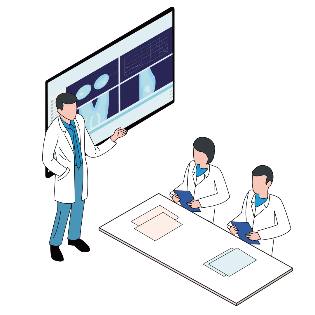
Many people imagine radiotherapy as simply “switch the machine on and shine it at the tumour”. Proton therapy could not be further from that. It is closer to haute couture: no two patients share the same plan.
Individualised proton therapy is a systematic, multidisciplinary undertaking, and the medical physicist plays the role of the designer. While the treatment plan is built for a patient, the physicist uses individualised field arrangement, dose optimisation and adaptive adjustment to strike the tumour precisely while protecting the surrounding normal organs as far as possible.
01 — First Define the Target Volume, Then Set the Dose Objectives
Target volume delineation
Clinical physicians use the planning CT — together with the patient's history, pathology and diagnosis — to define the tumour target volumes (GTV, CTV) and the organs at risk (OARs) nearby that need focused protection.
Dose constraints
Physicists and physicians set dosimetric objectives together, ensuring the lesion sits inside the high-dose region while the dose to normal tissue and organs at risk stays strictly below their tolerance thresholds. In one sentence: give the full dose where it must be hit, and hold the line where it must not.
02 — The Physicist's Core Work: Designing the “Bespoke Plan”
1) Field selection: no templates, every case different
The physicist weighs the realities of each case — the characteristics of the tissue the beam crosses to reach the tumour, the effect of potential motion and setup error on the dose, and the impact of the high biological effectiveness at the end of the field on organs at risk — and selects the field angles best suited to that patient.
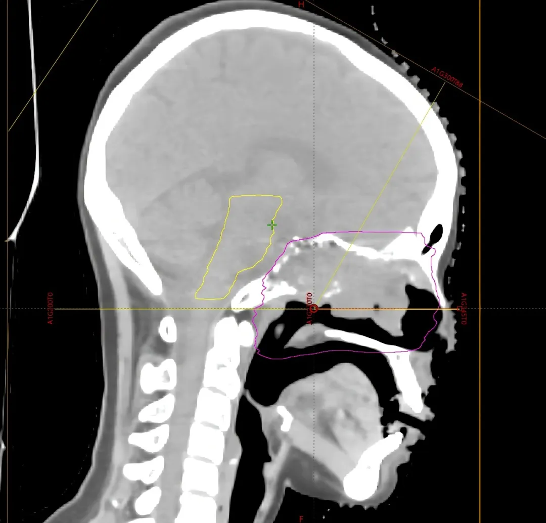
Take the skull-base tumour below. Because the target sits close to critical structures such as the optic nerve and brainstem, a conventional field would cross the optic nerve and its distal edge would land near the brainstem. Adding a non-coplanar field changes the picture: the beam direction runs parallel to the brainstem, bringing the brainstem dose down as far as it possibly can.
2) Beam and dose iterative optimisation — making the proton beam “stop” exactly where it should
The physicist exploits the Bragg peak, the physical signature of the proton beam, adjusting beam energy, position and intensity so that the high-dose region comes to rest precisely inside tumours at different depths: the dose falls rapidly to zero beyond the tumour. For special cases — patients carrying a breast implant, or respiratory movement of the lung — the physicist also obtains detailed material parameters or introduces a gated breathing feedback system, optimising the plan specifically so that the dose calculation is accurate and the dose distribution is as good as it can be.
03 — From Pre-Treatment Verification to In-Treatment Adjustment: Full-Course Dynamic Management
1) Before treatment: run a full “dress rehearsal”
Dose verification. Before formal treatment begins, the physicist reproduces the real treatment scenario end to end, carrying out a comprehensive dose check for every patient, every plan and every irradiation angle, so that what was planned matches what will actually be delivered. This step is like a dress rehearsal before opening night — treatment only starts with peace of mind once every element lines up.
2) During treatment: keep “fine-tuning”, i.e. adaptive planning
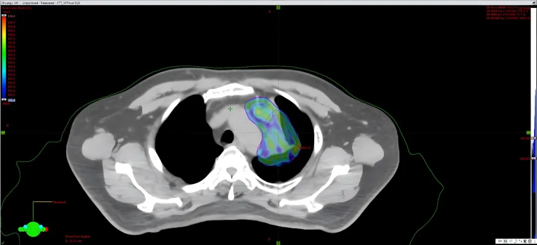
If during treatment the patient's weight changes, the tumour shrinks, or other anatomical structures change, the physicist and physician assess the consequences promptly and adjust the plan as needed (adaptive planning), so that the whole course stays precise and safe, with better dose delivery and better outcomes.
The lung cancer patient below is such a case. With marked tumour shrinkage during treatment, adaptive planning had to be performed promptly to bring down the dose to normal tissue while continuing to strike the tumour accurately.
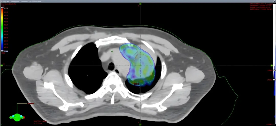
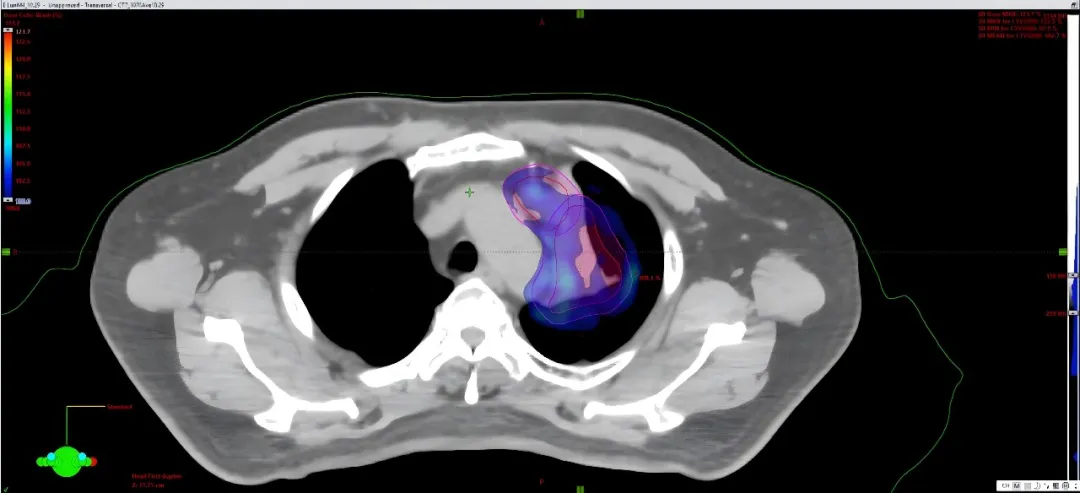
Proton therapy planning is not a mechanical “deliver the plan”. It requires the physicist to design the most suitable individualised plan according to the actual situation of each and every patient, and to keep monitoring and adjusting throughout the whole course of treatment.
This “one patient, one plan” precision design, combined with full-time monitoring and adjustment, is what maximises both the efficacy and the safety of proton therapy.
Written by Liu Kai, Medical Physics, Guangzhou Concord Cancer Center.